
RIES
Research Institute for Electronic Science, Hokkaido University
北海道大学
電子科学研究所
LAST UPDATE 2017/02/25
-
研究者氏名
Researcher Name西野浩史 Hiroshi NISHINO
助教 Assistant Professor -
所属
Affiliation北海道大学 電子科学研究所
人間数理研究分野
Research Institute for Electronic Science, Hokkaido University
Mathematical Modeling Lab. -
研究キーワード
Research Keywords聴覚器
微小センサー
昆虫
生物模倣
Auditory sense organ
Microsensor
Insects
Biomimetics
- 研究テーマ
Research Subject -
昆虫聴覚センサーの設計デザインの解明
Technical implementation of bauplan and material of insect auditory organ into microsensors
研究の背景 Background of the Research
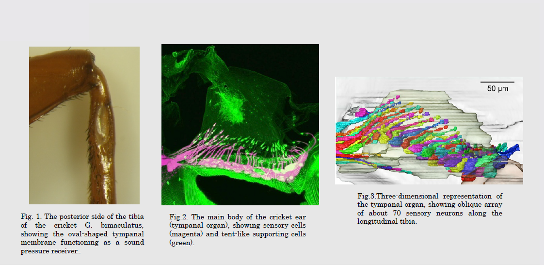
昆虫は長い進化の過程で体サイズに見合う微小センサーを生み出してきました。たとえば、コオロギの聴覚器(鼓膜器官)は我々の蝸牛の1/200のサイズにもかかわらず、1〜50kHZという広い可聴域を持ち、0.2nmの鼓膜の変位に応じることが知られています。近年の生物模倣の関心への高まりとともに昆虫のセンサーの設計原理を微小センサーの開発に応用しようとする機運が高まりつつあります。
Insects have been equipped with minute sensors through more than 60 million years evolution. For instance, the auditory organ (the tympanal organ) in a field cricket is 1/200 in size compared to the human cochlea. Nevertheless it achieves broad audible range (1-50 kHZ) and hyper-acute sensitivity (responding toto only 0.2 nm displacement of the tympanum). Concurrent with the accelerating attention of ecofriendly technology and biomimetics, insect mechanoreceptors draw growing attention as sensor models suitable for implementing their design principles into development of minute sensors of low energy cost, such as hearing aids and stretch detectors.
研究の目標 Research Objective
昆虫の聴覚器のインターフェースは我々と同じ鼓膜で、感覚細胞で発現するイオンチャネルも我々の内有毛細胞と同様にTRPチャネルです。ところが、伝音経路やこれを構成するマテリアルは我々の内耳とは大きく異なっています。伝音経路のinvivo粘弾性計測や構成成分の化学分析により、ヒトの蝸牛モデルと比肩する昆虫独自の聴覚受容モデルを作成することを目標とします。
Insects and mammalian ears share the tympanic membrane for efficient sound detection and TRP channels that convert mechanical distortion to electrical potential in sensory cells. However, sound transmission pathways interconnecting the tympanic membrane with sensory cells and materials transmitting sound energy in insects largely differ from our ears. Our ultimate goal is to simulate the auditory system comparable to the known cochlea model in humans. We address this subject using in vivo viscoelasticity measurements of the auditory organ and analysis of its chemical composition.
研究図Figures
論文発表 / Publications
昆虫と自然、印刷中(2015), 生物模倣技術と新材料・新製品開発への応用、生物の五感に学ぶ機能とその製品開発への応用、技術情報協会、第 2 章第 9 節, P208〜213 (2014)
研究者連絡先 / HP
- nishino
 es.hokudai.ac.jp
 - http://www.es.hokudai.ac.jp/labo/nishino/