IMRAM
Institute of Multidisciplinary Research for Advanced Materials, Tohoku University
東北大学
多元物質科学研究所

LAST UPDATE 2025/06/02
-
研究者氏名
Researcher Name百生敦 Atsushi MOMOSE
教授 Professor -
所属
Affiliation東北大学 多元物質科学研究所
計測研究部門量子ビーム計測分野
Institute of Multidisciplinary Research for Advanced Materials, Tohoku University
Division of Measurements,Quantum Beam Measurements -
研究キーワード
Research KeywordsX線光学
X線イメージング
位相コントラスト
X-ray optics
X-ray imaging
Phase contrast
- 研究テーマ
Research Subject -
X線位相イメージング技術の開拓と応用
Development and applications of X-ray phase imaging
研究の背景 Background of the Research
X線による透視撮影は、非破壊検査や医用画像診断、あるいは、学術用途のX線顕微鏡に至るまで、幅広い分野で活用されている。いずれの場合もX線の吸収コントラストで画像が形成されるのが一般的で、それゆえに軽元素からなる物質(高分子材料や生体軟組織など)に対する感度不足は原理的性質(欠点)として感受されてきた。これに対し、物質によるX線の位相シフトに基づく位相コントラストを使えば、この限界を克服できる。
X-ray radiography is used in a wide range of fields, from non-destructive testing and medical imaging diagnosis to X-ray microscopes for academic purposes. In all cases, images are generally formed by the absorption contrast of X-rays, and therefore the lack of sensitivity to materials made of light elements (such as polymer materials and soft biological tissues) has been recognized as a fundamental property (disadvantage). However, this limitation can be overcome by using phase contrast based on the phase shift of X-rays by materials.
研究の目標 Research Objective
我々は、X線透過格子を用いるX線位相イメージングを研究しており、実験室X線源を用いた装置化も可能となっている。X線位相イメージングのための光学系、および、それに用いるX線光学素子に関する研究を深化させ、X線位相情報を利用することの利点(軽元素材料において、原理的に約千倍の感度)を最大限に実現する光学システムの構築を果たし、産業界との協奏による実用化を押し進める。
We have pioneered X-ray phase imaging using X-ray transmission gratings, which has made it possible to develop devices using laboratory X-ray sources. We will deepen our research on optical systems for X-ray phase imaging and the X-ray optical elements used therein, and will build imaging systems that maximize the benefits of using X-ray phase information (in principle, approximately 1,000 times the sensitivity for light element materials), and will work with industry to promote practical application.
研究図Figures

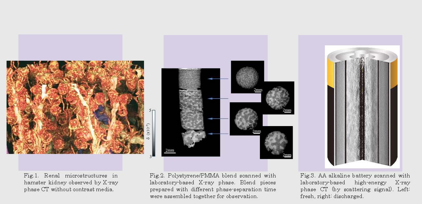
Fig.2. X-ray phase CT obtained for auditory ossicle of mouse (in collaboration with Prof. K. Matsuo, Keio Univ.).
Fig.3. in situ X-ray phase imaging (scattering image) of polymers under tensile test.
Fig.4. High-energy X-ray phase CT obtained for a tube battery.
論文発表 / Publications
Nat. Med. 2, 473 (1996). Jpn. J. Appl. Phys. 42, L866 (2003). Kid. Int. 75, 945 (2009). Opt. Express 19, 8423 (2011). Phil. Trans. R. Soc. A, 372, 2013023 (2014). Bone 84, 279 (2016). Macromolecules 38, 7197 (2005). Optica 6, 1012 (2019). Sci. Rep. 7, 6711 (2017). Proc. SPIE 11840, 118400K (2021).
研究者連絡先 / HP
- atsushi.momose.c2
 tohoku.ac.jp
 - https://www2.tagen.tohoku.ac.jp/lab/momose/