IMCE
Institute for Materials Chemistry and Engineering, Kyushu University
九州大学
先導物質化学研究所

LAST UPDATE 2017/02/25
-
研究者氏名
Researcher Name玉田薫 Kaoru TAMADA
教授 Professor -
所属
Affiliation九州大学 先導物質化学研究所
物質基盤化学部門・ナノ界面物性分野
Institute for Materials Chemistry and Engineering, Kyushu University
Division of Fundamental Organic Chemistry -
研究キーワード
Research Keywords表面プラズモン共鳴
ナノ粒子
バイオセンシング/バイオイメージング
Surface Plasmon Resonance
Nanoparticles
Bio-sensing / Bio-imaging
- 研究テーマ
Research Subject -
ナノ材料の多次元自己組織化とその応用
Multidimensionally self-assembled metallic nanoparticles and their applications
研究の背景 Background of the Research
金属ナノ粒子は、局在表面プラズモン共鳴により特定の波長の光をナノ粒子近傍に強く閉じ込めることができます。我々は、粒径が揃った金属ナノ粒子を気水界面に展開することで、金属ナノ粒子からなる二次元自己組織化膜(プラズモニックナノシート)を作製し、その光学物性および電気特性について調べると共に、バイオセンシング・バイオイメージングなどへの応用を目指しています。
Metal nanoparticles can confine the light at nanointerface. We fabricate a 2D nanosheet composed of self-assembled metal nanoparticles at air-water interface to investigate their fundamental optical and electrical properties (‘plasmonic nanosheet’). This study goes toward a unique application in the field of bio-sensing and bio-imaging.
研究の目標 Research Objective
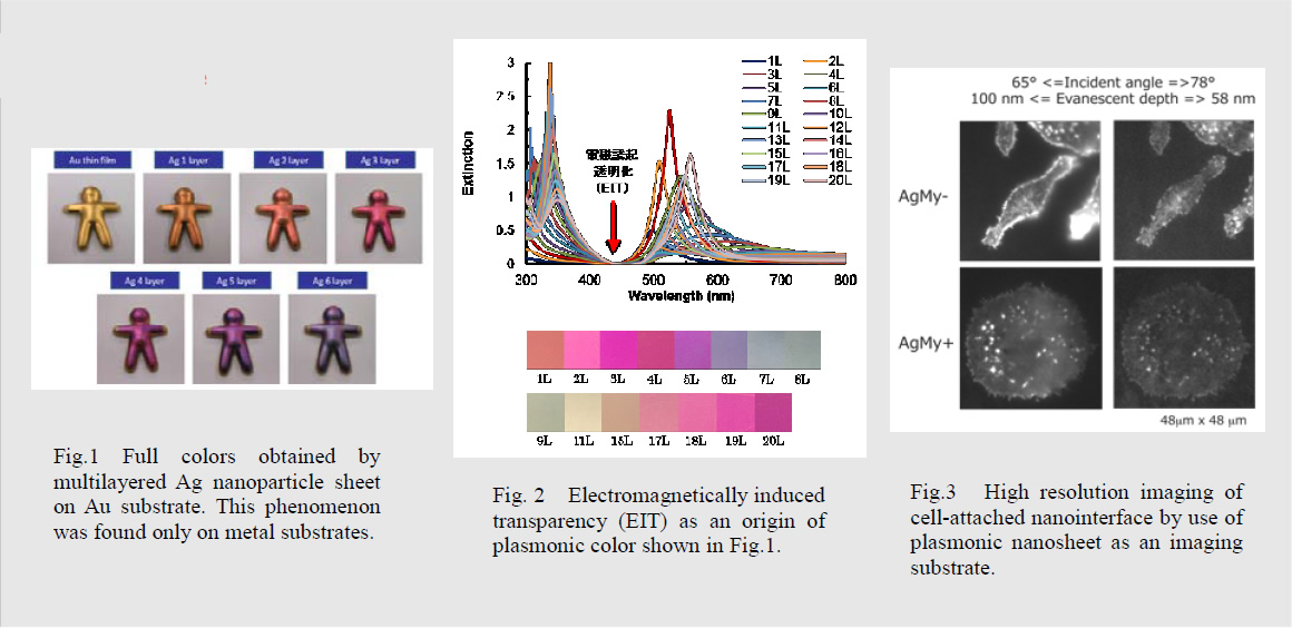
金属ナノ微粒子の多次元自己組織化構造および複雑系における局在プラズモンの協同励起現象を解明し、これを高感度バイオセンシングや高分解能バイオイメージングに応用し、ライフ・イノベーションを推進します。例えば、金属薄膜上にプラズモニックナノシートを積層すると積層数によって鮮やかな呈色変化がみられますが、これはこの膜のメタマテリアル的性質に起因するもので、この現象を利用した目視型フルカラーセンサーの開発を進めています。
The main object of our research is to clarify the collective excitation of localized surface plasmons at multidimensionally assembled structures or more complex systems for life-innovative study. For example, multi-layered plasmonic nanosheet exhibits a unique metallic full-color due to their optical property as metamaterials. We propose a new type of biosensors for eye-detection by use of this phenomenon.
研究図Figures

論文発表 / Publications
K. Okamoto, et al, Sci. Rep., in press (2016). R. Degawa et al, Langmuir, 32, 8154-8162 (2016). D. Tanaka et al, Nanoscale. 7, 15310-15320 (2015).
S. Shinohara, et al, Phys. Chem. Chem. Phys. 17, 18606-18612 (2015).
研究者連絡先 / HP
- tamada
 ms.ifoc.kyushu-u.ac.jp
 - http://www.cm.kyushu-u.ac.jp/ktamada/