IMRAM
Institute of Multidisciplinary Research for Advanced Materials, Tohoku University
東北大学
多元物質科学研究所

LAST UPDATE 2021/05/06
-
研究者氏名
Researcher Name江島丈雄 Takeo EJIMA
准教授 Associate Professor -
所属
Affiliation東北大学 多元物質科学研究所
計測研究部門 放射光ナノ構造可視化研究分野
Institute of Multidisciplinary Research for Advanced Materials, Tohoku University
Division of Measurements, Synchrotron Radiation Soft X-ray Microscopy -
研究キーワード
Research Keywords軟Ⅹ線光学
軟Ⅹ線相関顕微法
「水の窓」「炭素の窓」波長域
軟Ⅹ線利用計測法
Soft X-ray Optics
Soft X-ray Correlation Spectromicroscopy
Water Window, Carbon Window
Application of Soft X-ray optics
- 研究テーマ
Research Subject -
軟Ⅹ線相関顕微法の開発とその生物細胞観察応用
Development of Soft X-ray Correlation Spectromicroscopy applying to Bio-cells
研究の背景 Background of the Research
軟Ⅹ線は、物質を構成する元素内の電子と強く相互作用し、可視光よりも1桁以上波長が短いという2つの特徴を持ちます。近年開発された高輝度軟Ⅹ線源を利用すると、この2つの特徴を生かした分光顕微計測が可能です。このような分光顕微計測法は、生物細胞のように多くの微小構造から成りその機能がよく分からない物質を計測するために必要とされています。
The light in soft X-ray wavelength region has two features; strong interaction with electrons of elements in materials, and short wavelength that is one or more order shorter than the one of visible light. Fine spectromicroscopy measurement using these two features will be practicable by the use of high brilliance light source developed recently. The spectromicroscopy measurement enables us to clarify functions of complex materials such as small organelles in bio-cells.
研究の目標 Research Objective
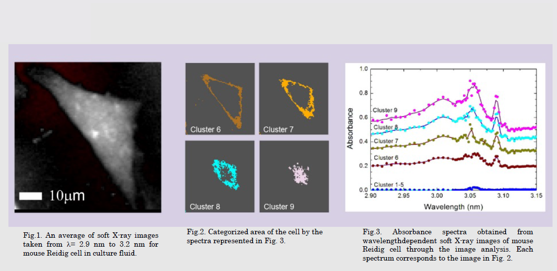
細胞の軟Ⅹ線像の波長依存性を測定すると、微小構造ごとに波長依存性が異なるため、波長により異なった形状に見えます。細胞中の微小細胞内小器官の構造と機能を明らかするために、高輝度軟Ⅹ線源と軟Ⅹ線光学を利用した顕微鏡により撮像した、波長依存の画像の波長の持つ情報と構造の持つ情報を対応させる相関顕微計測法の開発を行っています。
Small organelles in soft X-ray images show different shape according to the wavelength because wavelength dependence of the organelles is different. Correlation between soft X-ray spectra and shapes observed in the soft X-ray images will clarify the fine structures and the functions of the small organelles. Spectromicroscopic methodology to measure the correlation is investigating by use of soft X-ray optics and high brilliance soft X-ray light source.
研究図Figures

論文発表 / Publications
江島丈雄、加道雅孝「「水の窓」波長域の軟Ⅹ線顕微鏡による生物細胞観測」O plus E 36(3), (2014) 296. 江島丈雄、「軟 X 線による超解像度光学顕微鏡の開発」、光アライアンス、23(4) (2012) pp. 1-4
研究者連絡先 / HP
- takeo.ejima.e7
 tohoku.ac.jp
 - https://www2.tagen.tohoku.ac.jp/lab/takata/html/