IMRAM
Institute of Multidisciplinary Research for Advanced Materials, Tohoku University
東北大学
多元物質科学研究所
LAST UPDATE 2021/05/05
-
研究者氏名
Researcher Name髙橋幸生 Yukio TAKAHASHI
教授 Professor -
所属
Professional Affiliation東北大学多元物質科学研究所
無機材料研究部門 放射光可視化情報計測研究分野
Institute of Multidisciplinary Research for Advanced Materials, Tohoku University
Division of Inorganic Material Research, Synchrotron Radiation Microscopy and Informatics -
研究キーワード
Research KeywordsコヒーレントX線光学
放射光
可視化計測
情報科学
Coherent X-ray Optics
Synchrotron Radiation
Visualization Measurement
Informatics
- 研究テーマ
Research Subject -
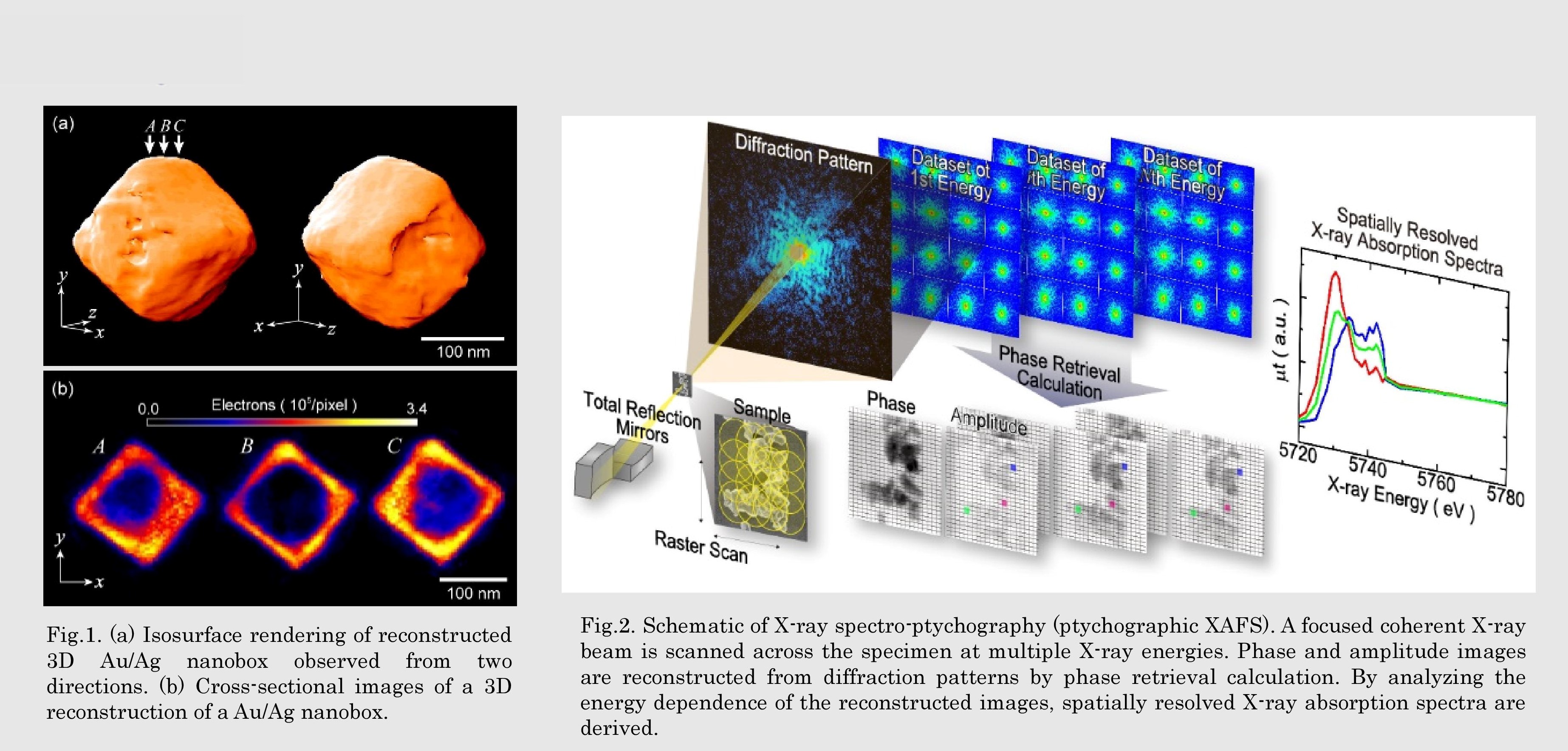
コヒーレントX線によるナノ構造可視化法の開発とその応用展開
Development and application of nano imaging methods using coherent X-ray
研究の背景 Background
実用材料の多くは、ナノからマイクロメートルまでの空間階層構造を有する複雑系です。したがって、新材料を設計・開発する際、ナノ・メソスケールでの微細構造と機能の相関を解明することが重要です。放射光のコヒーレント成分を利活用したコヒーレントX線回折イメージングは、X線領域で未踏であったナノスケールでの構造可視化を実現する次世代の可視化計測法であり、実用材料の空間階層構造を解析するツールとして注目されています。
Many practical materials are heterogeneous complex systems with hierarchical structures from nanometer to micrometer scale. It is therefore important to understand correlation between the fine structures and the function at nano-meso scale to create new functional materials. Coherent X-ray diffraction imaging is a promising method for visualizing the structures inside bulk materials at the nanoscale
研究の目標 Outcome
先進的X線光学技術を駆使することで、物質のナノ・メソスケールでの構造・元素・化学状態を3次元的に可視化する新規コヒーレントX線回折イメージング法を開発します。さらに、情報科学を活用することで実用材料の構造-機能相関に関する特徴的な情報を抽出する基盤を構築することを目指します。
Our mission is the development of next-generation synchrotron radiation microscopy /spectroscopy methods using novel X-ray optics. Finally, we will create the platform to visualize the function of practical materials using informatics.
研究図Research Figure
文献 / Publications
Nano Lett., 10, 1922 (2010). Nano Lett., 13, 6028 (2013). Phys. Rev. Lett., 112, 053903 (2014). Angew. Chem. Int. Ed., 130, 1490 (2018). Communications Chemistry, 2, 50 (2019).
研究者HP
- ytakahashi
 tohoku.ac.jp
 - http://www2.tagen.tohoku.ac.jp/lab/takahashi-y/html/index.html