
RIES
Research Institute for Electronic Science, Hokkaido University
北海道大学
電子科学研究所

LAST UPDATE 2022/01/14
-
研究者氏名
Researcher Name鈴木明大 Akihiro SUZUKI
准教授 Associate Professor -
所属
Affiliation北海道大学 電子科学研究所
光科学研究部門 コヒーレント光研究分野
Research Institute for Electronic Science, Hokkaido University
Laboratory of Coherent X-ray Optics, Section of Photonics and Optical Science -
研究キーワード
Research KeywordsX線自由電子レーザー
コヒーレント回折
ナノイメージング
微細加工
X-ray free-electron laser (XFEL)
Coherent diffraction
Nanoimaging
Microfabrication
- 研究テーマ
Research Subject -
生物・材料試料のコヒーレントX線イメージング
Coherent X-ray imaging of biological and materials science samples
研究の背景 Background of the Research
X線光源はもちろん、微細加工技術や計算機の発達に伴い、X線のコヒーレンスに基づいたイメージングの研究が、2000年ごろより世界中の放射光施設で活発化している。高い透過能などのX線の特徴を生かすことで、他のプローブではアクセスできない原子・ナノ構造の可視化が期待される。
With the development of microfabrication technologies and computing power as well as X-ray light sources, X-ray nanoimaging research based on coherence has been studied intensively in synchrotron radiation facilities across the world since around 2000. Utilizing the characteristics of X-rays such as penetration power, visualization of atomic and nanostructures that cannot be accessed by other probes is expected.
研究の目標 Research Objective
これまでに、微細加工技術で作製したマイクロ液体アレイを用いて、溶液中の金属ナノ粒子や生細胞のイメージングに成功してきた。今後は、超高密度X線レーザーを利用できるイメージングシステムを構築することで、生体超分子やタンパク質の単粒子イメージングに挑む。さらに、sub-10 nmでの磁気構造分析を目標に、X線磁気円二色性に基づいた磁気イメージングも展開する。
We have succeeded in nanoimaging metal nanoparticles and living cells in solution using micro-liquid cell arrays fabricated by microfabrication technology. From now on, we will challenge the single-particle imaging of biological supramolecules and proteins by developing an imaging system that can utilize ultra-high-density X-ray laser pulses. In addition, we will develop a magnetic imaging system based on X-ray magnetic circular dichroism to analyze magnetic structures at sub-10 nm.
研究図Figures
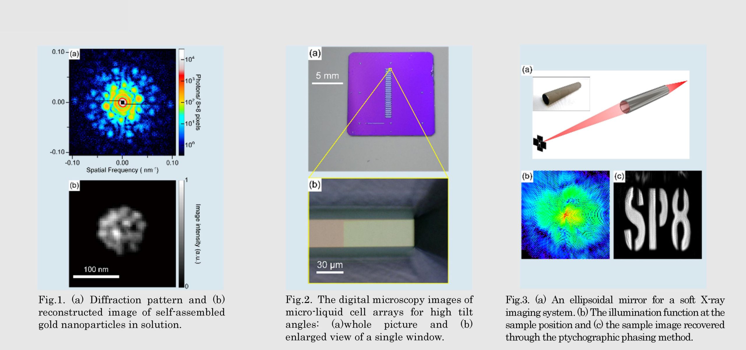
Fig.2. The digital microscopy images of micro-liquid cell arrays for high tilt angles: (a)whole picture and (b) enlarged view of a single window.
Fig.3. (a) An ellipsoidal mirror for a soft X-ray imaging system. (b) The illumination function at the sample position and (c) the sample image recovered through the ptychographic phasing method.
論文発表 / Publications
Rev. Sci. Instrum., 91, 083706 (2020). AIP Advances, 10, 055219 (2020). App. Phys. Lett., 116, 121102 (2020), Phys. Chem. Chem. Phys, 22, 2622 (2020).
研究者連絡先 / HP
- Akihiro.suzuki
 es.hokudai.ac.jp
 - https://researchmap.jp/akihiro.suzuki