
RIES
Research Institute for Electronic Science, Hokkaido University
北海道大学
電子科学研究所
LAST UPDATE 2017/02/25
-
研究者氏名
Researcher Name西野吉則 Yoshinori NISHINO
教授 Professor -
所属
Affiliation北海道大学 電子科学研究所
光科学研究部門・コヒーレント光研究分野
Research Institute for Electronic Science, Hokkaido University
Laboratory of Coherent X-ray Optics, Section of Photonics and Optical Science -
研究キーワード
Research KeywordsコヒーレントX線
放射光
X線自由電子レーザー
位相コントラストイメージング
Coherent X-rays
Synchrotron radiation (SR)
X-ray free-electron laser (XFEL)
Phase-contrast imaging
- 研究テーマ
Research Subject -
コヒーレント X 線を用いた物質深部のナノ構造解析
Analysis of nanostructures deep inside materials using coherent X-rays
研究の背景 Background of the Research
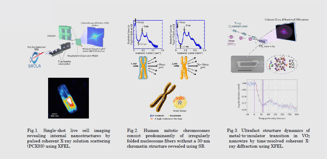
X線回折は、伝統的に、結晶試料に対する原子構造解析に威力を発揮してきた。さらに、コヒーレントX線による回折を用いることにより、例えば、細胞や細胞小器官など、結晶化できない試料に対しても、高空間分解能での構造解析への扉が開く。フェムト秒オーダーのパルス幅をもつX線自由電子レーザーを利用すると、高分解能での生物イメージングで問題となってきた試料の放射線による損傷の問題を初めて根源的に解決し、自然な状態にある生物試料をスナップショットイメージングできる。
Traditionally, x-ray diffraction has been powerfully used in atomic structure determination for crystalline samples. Coherent x-rays diffraction further opens up a new avenue for high-resolution structure analysis even for non-crystalline sample, such as cells or organelles. X-ray free-electron lasers with femtosecond pulse duration for the first time provide an opportunity to solve the radiation damage problem in high-resolution biological imaging, and enable us to take snapshots of biological samples under natural conditions.
研究の目標 Research Objective
放射光やX線自由電子レーザーなどの先端的短波長コヒーレント光源の特徴を最大限活かし、マクロな世界から原子の世界までをイメージングする基礎および応用研究を展開する。これは、我々にとって関心の対象となるマクロな機能を、原子・ナノ構造と結びつけて理解する上で極めて重要であり、生命科学から物質科学に至る幅広い科学分野で、新しい科学的知見を与えるブレークスルーをもたらすと期待する。
We take advantage of the coherence of synchrotron radiation (SR) and X-ray free-electron laser (XFEL) to develop new imaging techniques in a wide length scale ranging from macroscopic to atomic. We have been performing coherent X-ray imaging research to understand macroscopic properties from atomic-/nano-level, which will definitely lead to breakthroughs in a variety of scientific fields including both life and materials sciences.
研究図Figures
論文発表 / Publications
Nature Commum., 5, 3052 (2014). Nano Lett., 14, 2413(2014). Opt. Express, 21, 9267 (2013). Curr. Opin. Struct. Biol., 22, 670 (2012). EMBO J., 31, 1644 (2012). Appl. Phys. Express, 3, 102701 (2010). Nature Phys., 6, 122 (2010). Phys. Rev. Lett., 102, 018101 (2009). Proc. Natl. Acad. Sci. USA, 100, 110 (2003).
研究者連絡先 / HP
- yoshinori.nishino
 es.hokudai.ac.jp
 - http://cxo-www.es.hokudai.ac.jp/