IMRAM
Institute of Multidisciplinary Research for Advanced Materials, Tohoku University
東北大学
多元物質科学研究所

LAST UPDATE 2021/05/04
-
研究者氏名
Researcher Name百生敦 Atsushi MOMOSE
教授 Professor -
所属
Professional Affiliation東北大学多元物質科学研究所
計測研究部門 量子ビーム計測研究分野
Institute of Multidisciplinary Research for Advanced Materials, Tohoku University
Division of Measurements, Quantum Beam Measurements -
研究キーワード
Research KeywordsX線光学
X線イメージング
位相コントラスト
X-ray optics
X-ray imaging
Phase contrast
- 研究テーマ
Research Subject -
X線位相イメージング手法の開拓
Development of X-ray phase imaging
研究の背景 Background
X線の位相情報、あるいは、コヒーレンスの活用技術が大きく進歩し、X線イメージング分野は大きく進展しています。吸収コントラストに頼る従来のX線透視画像は医療や非破壊検査などに広く活用されていますが、X線位相コントラストの利用が広まることにより、従来法ではコントラストが付きづらい軽元素から成る高分子材料や生体軟組織などの物質が観察できるようになります。
The techniques for utilizing X-ray phase information or coherence have been developed remarkably, and the field of X-ray imaging is progressing well. Whereas conventional X-ray imaging methods relying on absorption contrast are widely used in medicine and non-destructive testing, X-ray phase-contrast method allows us to observe materials consisting of low-Z elements, such as polymers and biological soft tissues, which cannot be depicted with the conventional methods.
研究の目標 Outcome
X線光学を深く探求し、位相情報を活用するX線顕微鏡や断層撮影法(X線CT)などの先端画像計測技術の開拓を行っています。また、中性子線などの他の量子ビームを用いた位相イメージング研究にも活動を展開します。医用画像機器や非破壊検査装置への展開など、産業応用も視野に入れ、健康・安心・安全に関心の高い現代社会への貢献を目指します。
X-ray optics is extensively studied to develop advanced imaging methods, such as X-ray microscopy or tomography utilizing phase information. Phase imaging with other quantum beams, such as neutron beam, is also studied. Through these activities, contribution to the modern society with increasing attention to health, safe, and secure by developing apparatus for medical diagnosis and non-destructive testing in collaboration with industry.
研究図Research Figure

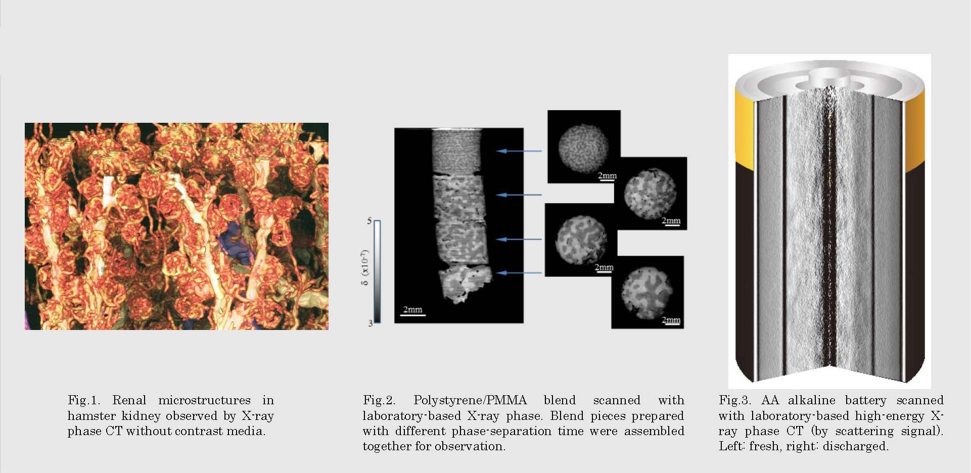
Fig.2. Polystyrene/PMMA blend scanned with laboratory-based X-ray phase. Blend pieces prepared with different phase-separation time were assembled together for observation.
Fig.3. AA alkaline battery scanned with laboratory-based high-energy X-ray phase CT (by scattering signal). Left: fresh, right: discharged.
文献 / Publications
Nat. Med. 2, 473 (1996). Jpn. J. Appl. Phys. 42, L866 (2003). Kid. Int. 75, 945 (2009). Opt. Express 19, 8423 (2011). Phil. Trans. R. Soc. A, 372, 2013023 (2014). Bone 84, 279 (2016). Macromolecules 38, 7197 (2005). Optica 6, 1012 (2019). Sci. Rep. 7, 6711 (2017). Proc. SPIE 11840, 118400K (2021).
研究者HP
- atsushi.momose.c2
 tohoku.ac.jp
 - http://mml.tagen.tohoku.ac.jp/