
RIES
Research Institute for Electronic Science, Hokkaido University
北海道大学
電子科学研究所
LAST UPDATE 2019/06/10
-
研究者氏名
Researcher Name田口敦清 Atsushi TAGUCHI
准教授 Associate Professor -
所属
Affiliation北海道大学 電子科学研究所
光科学研究部門・光システム物理研究分野
Research Institute for Electronic Science, Hokkaido University
Laboratory of Photo-System Physics, Photonics and Optical Science -
研究キーワード
Research Keywordsナノフォトニクス
プラズモニクス
ナノ分光イメージング
深紫外フォトニクス
Nanophotonics
Plasmonics
Nano Spectroscopy & Nano imaging
Photonics in Deep UV
- 研究テーマ
Research Subject -
紫外領域のナノフォトニクスの開拓
DUV nanophotonics
研究の背景 Background of the Research
紫外光はフォトンのエネルギーが高く、物質との強い相互作用が顕微鏡や分光分析、リソグラフィをはじめとする科学や産業の様々な用途に用いられています。その紫外光とプラズモニクスやナノフォトニクスの技術を組み合わせ、局在性や増強度を飛躍的に高めた新しい紫外技術を開拓しています。
Ultraviolet (UV) light has high photon energy and it strongly interacts with materials through the electronic transitions. The unique properties of UV light have been applied in a variety of scientific and industrial applications including microscopy, spectroscopy, lithography, material processing, and so on. We aim to extend the UV technology by combining with plasmonics and nanophotonics, exploring the frontier of UV technology that is realized by highly localized and enhanced UV light at nanoscale.
研究の目標 Research Objective
紫外光領域の光と物質との相互作用を活用した高解像度なイメージング技術や物質変換技術を開拓しています。特に、(1)紫外光を用いた生体イメージング技術の開発(Fig. 1)、(2)紫外プラズモニクス(Fig. 2)を中心に進めています。
We are developing high-resolution imaging technique using light-matter interactions in the UV region at nanoscale. Specifically, development of (1) bioimaging techniques using DUV light (Fig. 1) and (2) UV plasmonics (Fig. 2) are currently the main projects.
研究図Figures
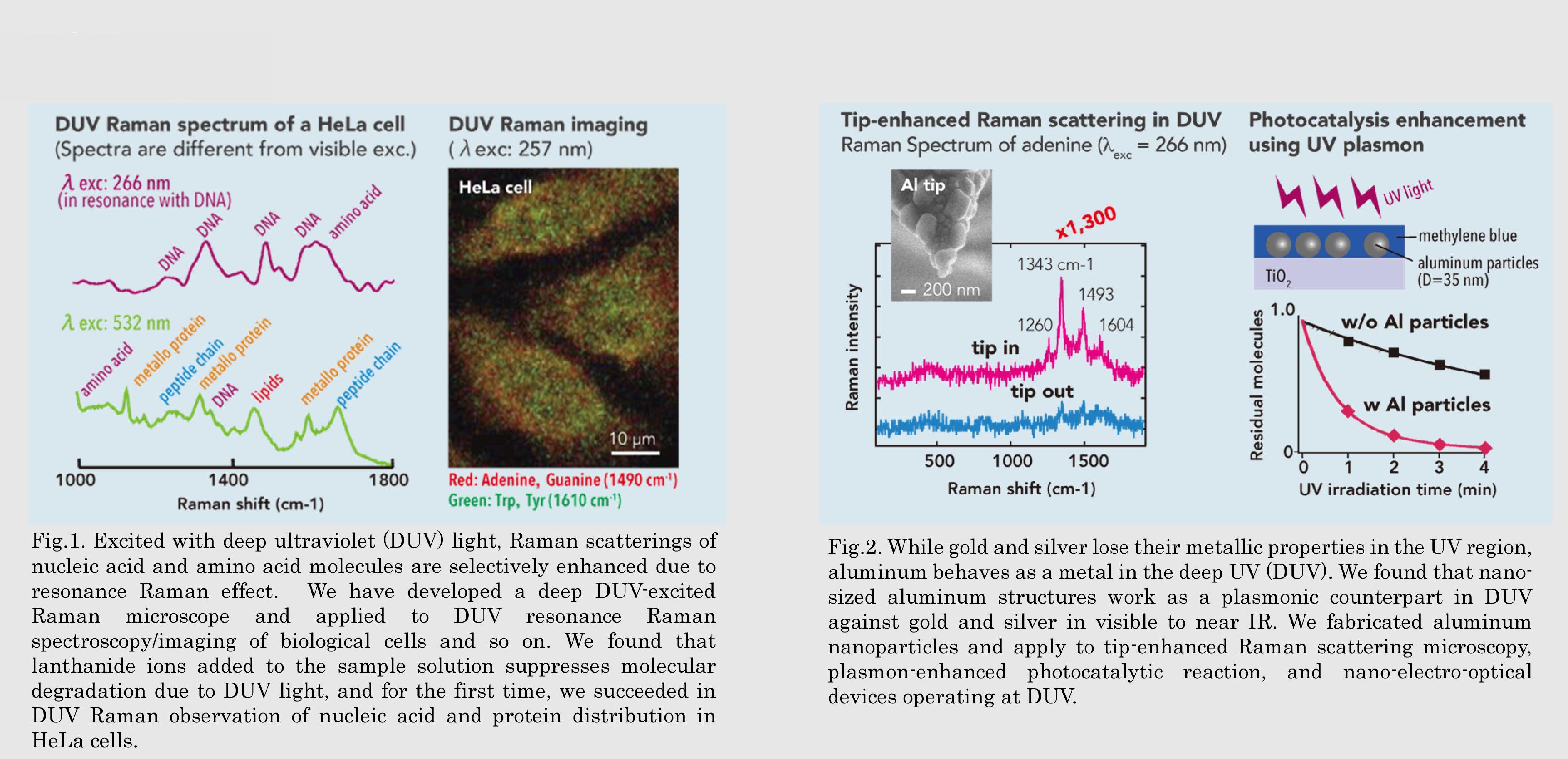
Fig.2. While gold and silver lose their metallic properties in the UV region, aluminum behaves as a metal in the deep UV (DUV). We found that nano-sized aluminum structures work as a plasmonic counterpart in DUV against gold and silver in visible to near IR. We fabricated aluminum nanoparticles and apply to tip-enhanced Raman scattering microscopy, plasmon-enhanced photocatalytic reaction, and nano-electro-optical devices operating at DUV.
論文発表 / Publications
J. Raman Spectrosc. 40, 1324 (2009); Appl. Phys. Lett. 101, 081110 (2012); Appl. Phys. Lett. 104, 061108 (2014); Nanoscale 7, 17424 (2015); Chem. Rev. 117, 4983 (2017); Chem. Soc. Rev.46, 4077 (2017); Adv. Opt. Mater. 7,1801099 (2019); APL Photonics 4, 021301 (2019).
研究者連絡先 / HP
- taguchi
 es.hokudai.ac.jp
 - http://optsys.es.hokudai.ac.jp/taguchi/