
IMRAM
Institute of Multidisciplinary Research for Advanced Materials, Tohoku University
東北大学
多元物質科学研究所
LAST UPDATE 2021/05/01
-
研究者氏名
Researcher Name水上進 Shin MIZUKAMI
教授 Professor -
所属
Affiliation東北大学 多元物質科学研究所
有機・生命科学研究部門 細胞機能分子化学研究分野
Institute of Multidisciplinary Research for Advanced Materials, Tohoku University
Division of Organic- and Bio-materials Research, Cell Functional Molecular Chemistry -
研究キーワード
Research Keywordsバイオイメージング
細胞機能制御
光機能性分子
Bioimaging
Regulation of cellular functions
Photofunctional molecules
- 研究テーマ
Research Subject -
機能性分子設計による細胞機能の可視化と制御
Imaging and regulation of cellular functions with functional molecule design
研究の背景 Background of the Research
生体内では多くの生体分子が相互作用することで、多様な機能を発現しています。生物を正確に理解するには、生体分子の挙動や機能を他の分子との相互作用が保たれた状態、すなわち生きた状態で調べることが重要になります。近年、化学的なアプローチに基づいて、生命機能を調べるChemicalbiologyと呼ばれる学問が注目を集めています。
In a living body or cell, various biomolecules function by interacting with other molecules. To understand the real biological functions, it is essential to investigate the activities or behaviors of the target biomolecules in living systems, where all of these interactions with other biomolecules are maintained. Recently, a new research field “Chemical biology”, which is a study of biomolecular functions on the basis of chemical approaches has been attracting increasing attention.
研究の目標 Research Objective
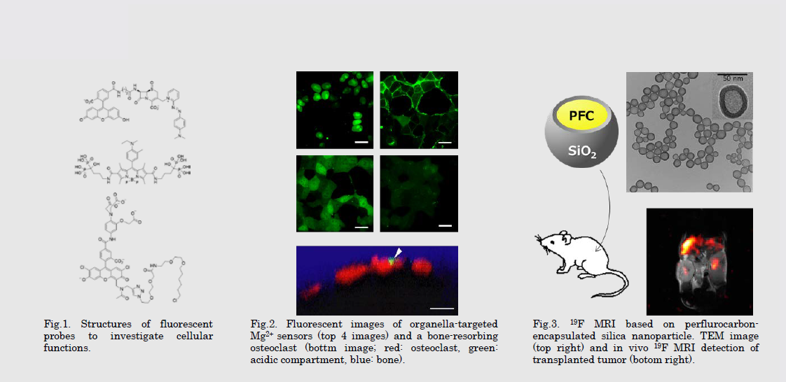
有機化学・高分子化学・蛋白質化学等に基づいて新たな機能性分子を設計・合成し、生体機能の探索技術を開発しています。具体的には、酵素活性や細胞内シグナル伝達などの生体機能を選択的に可視化する蛍光プローブ、ケージド化合物/フォトクロミック化合物を用いた蛋白質活性の光制御技術などを開発し、最先端のイメージング技術と組み合わせることにより、生きた状態における生体分子の機能や疾患機構の本質に迫ります。
Using multidisciplinary chemistry such as organic chemistry, macromolecular chemistry, and protein chemistry, we design and synthesize functional molecules, and apply them to image or regulate behaviors or functions of target biomolecules. By combining these functional compounds with the latest imaging technologies, we aim to clarify essential mechanisms of life and various diseases.
研究図Figures
論文発表 / Publications
Nat. Chem. Biol., 12, 579 (2016). Angew. Chem. Int. Ed., 524, 1007 (2015). Angew. Chem. Int. Ed. 53, 1008 (2014). Acc. Chem. Res. 47, 247 (2014). J. Am. Chem. Soc. 134, 1623 (2012). J. Am. Chem. Soc. 133, 17772 (2011). J. Am. Chem. Soc. 132, 9524 (2010).
研究者連絡先 / HP
- s-mizu
 tagen.tohoku.ac.jp
 - http://www2.tagen.tohoku.ac.jp/lab/mizukami/